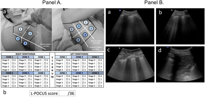
Fig 1. Lung point-of-care ultrasonography method (L-POCUS) and examples of four ultrasound aeration stages.
(Panel A). (a). Twelve chest areas of investigation following BLUE-PLUS Protocol: zone 1: upper anterior chest wall; zone 2: lower anterior chest wall; zone 3: upper lateral chest wall; zone 4: lower lateral chest wall; zone 5: upper posterolateral chest wall; zone 6: lower posterolateral chest wall. (b) L-POCUS score grid: Each zone was examined to establish which of four ultrasound parenchymal aeration stages it exhibited, and points are assigned to them according to their severity. Stage 0 or normal aeration (0 point): Lung sliding sign associated with respiratory movement of less than 3 B lines; Stage 1 or moderate loss of lung aeration (1 point): a clear number of multiple visible B-lines with horizontal spacing between adjacent B lines ≤ 7 mm (B1 lines); Stage 2 or severe loss of lung aeration (2 points): multiple B lines fused together that were difficult to count with horizontal spacing between adjacent B lines ≤ 3 mm, including “white lung”; and Stage 3 or pulmonary consolidation (3 points): hyperechoic lung tissue, accompanied by dynamic air bronchogram. (Panel B). (a) Stage 0 or normal aeration; (b) Stage 1 or moderate loss of lung aeration; (c) Stage 2 or severe loss of lung aeration; (d) Stage 3 or pulmonary consolidation.

