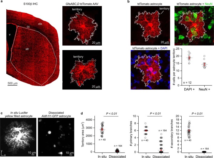
Extended Data Fig. 1. Morphologically complex striatal astrocytes lose their complexity upon dissociation.
a. Representative image of S100β immunostaining in a mouse coronal section containing the striatum. Subpanels show representative striatal astrocytes sparsely expressing cytosolic tdTomato fluorescent protein after GfaABC1D tdTomato AAV microinjection. The experiment was replicated five times. b. Striatal astrocyte sparsely labeled with tdTomato and co-stained with nuclear marker DAPI and neuronal marker NeuN. Quantification shows DAPI+ nuclei and NeuN+ cell bodies found in a single tdTomato+ astrocyte’s territory. Mean and SEM are shown; (n = 12 tdTomato+ cells per group from 4 animals). c. In situ Lucifer yellow iontophoretically filled striatal astrocyte from 8-week-old mice. Right panel shows GFP+ striatal astrocytes after dissociation for fluorescence activated cell sorting (FACS) from 8-week-old mice. d. Quantification of territory area, number of primary branches, and number of secondary branches showed that dissociated astrocytes display decreased cellular complexity. Mean and SEM are shown; (n = 40 cells from 15 mice for Lucifer yellow and n = 164 cells from 8 mice for Aldh1l1-GFP, respectively; territory area and number of primary branches: two-tailed student’s unpaired t-test, P = 0.0002; number of secondary branches: two-tailed one-sample t-test, P = 0.003).

