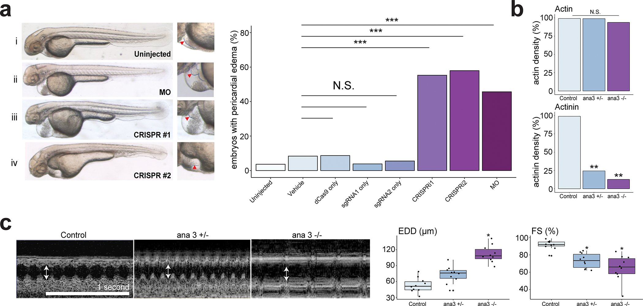
Figure 3. RTTN is an evolutionarily conserved causal gene for iDCM.

a, Left, representative images of embryos at 48 hpf. (i) uninjected embryo; (ii) embryo injected with translational blocking morpholino (MO); embryos injected with CRISPRi #1 (iii) and #2 (iv). Right, corresponding enlarged images of the heart. Arrowhead, outline of heart. Incidence of pericardial edema with impaired tail circulation in 48-hpf embryos. Results n ≥ 101 embryos from n ≥ 7 biological replicates using embryos from n ≥ 2 breeding pairs. Vehicle was 0.08% phenol red + PBS in ultrapure water. ***p <0.00017 by Fisher’s exact test with Bonferroni Correction; N.S. = not significant. b, Quantification of immunofluorescent imaging of actin and actinin in adult heart (segment A4) from control (w1118), ana3+/− and ana3−/− flies. Statistical results of normalized cardiac fiber density (N = 10). * P <0.05; ** P <0.01. c, OCT images for heart of control, ana3+/− and ana3−/− flies. Arrowheads indicate\ the end-diastolic diameter (EDD). Statistical analysis for EDD (μm) and percent fractional shortening (FS) obtained from the optimal computed tomography data. Each datapoint represents the average of measurements from three heartbeats randomly selected within a two-second time frame for each fly. Center line = median; whiskers = 1.5IQR for each genotype. *P <0.05.
