
Figure 10:
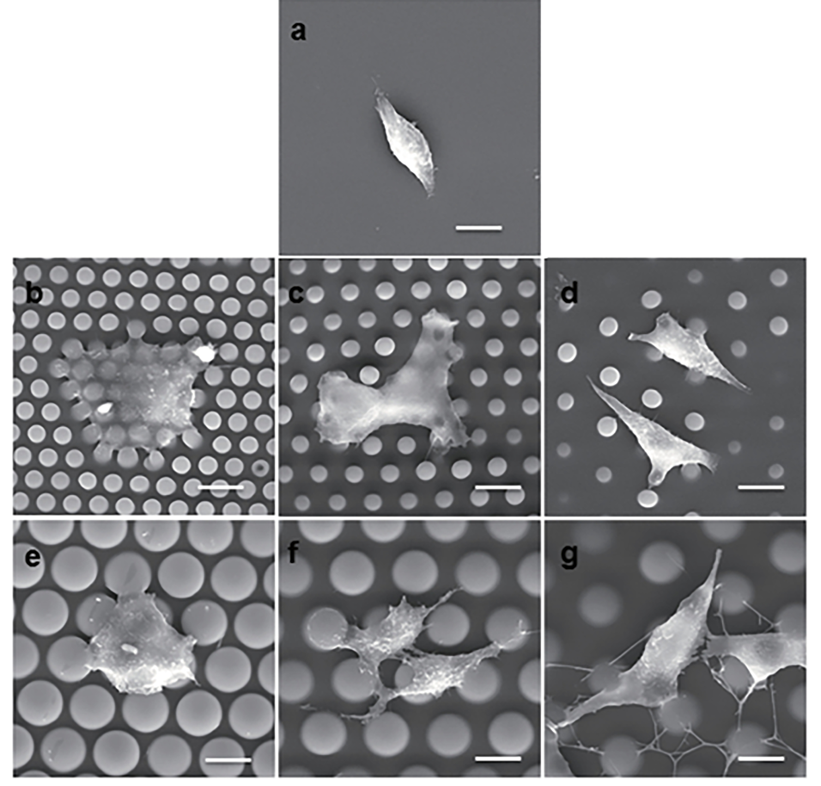
Cellular morphology on micropillar arrays and planar. A: Micropillar-induced cellular morphogenesis. (a) An SEM micrograph shows A549 cells on the flat PDMS substrate. (b–g) SEM micrographs show A549 cells on the 4–2 μm (b), 4–4 μm (c), 4–7 μm (d), 10–2 μm (e), 10–4 μm (f), and 10–7 μm (g) micropillar arrayed substrates. Scale bar =10 μm. Figure reproduced with permission from Xu et al. 220.