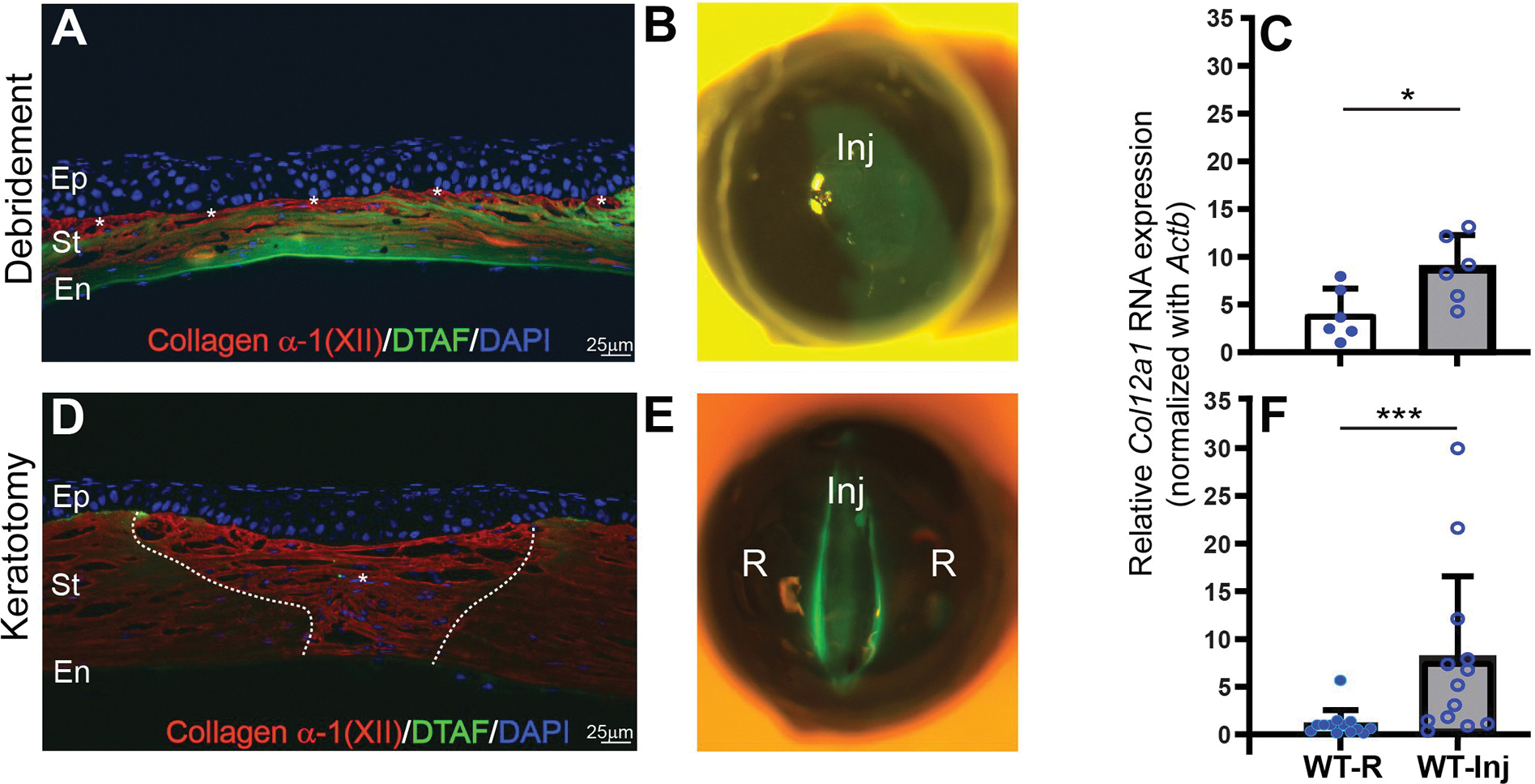
Figure 1: Expression of collagen XII following trauma three weeks after injury in two different types of injury.

(A) A model of deep burr injury in the stroma with loss of stromal tissue shows expression of collagen XII in the surface of the cornea, white asterisks. (B) Slit-lamp microphotograph demonstrates area of burr injury demarcated by DTAF. (C) Quantitative PCR quantification shows statistically significant expression of Col12a1 mRNA transcripts compared to control contralateral uninjured eye; Wilcoxon paired test. (D) A lineal full thickness keratotomy injury with expression of collagen XII in the newly regenerated wound. White asterisk shows newly regenerated tissue expressing collagen XII. (E) Slit-lamp microphotograph demonstrates area of full thickness keratotomy injury demarcated by DTAF. New tissue regeneration and remodeling is located within DTAF area. (F) Quantitative PCR quantification shows statistically significant expression of Col12a1 mRNA transcripts compared to control uninjured area of the same eye. Wilcoxon paired test. DTAF: (5-(4,6-dichlorotriazinyl) aminofluorescein). Inj: area of injury, R: area outside injury. *P < 0.05; ***P < 0.005. Bar 25μm.
