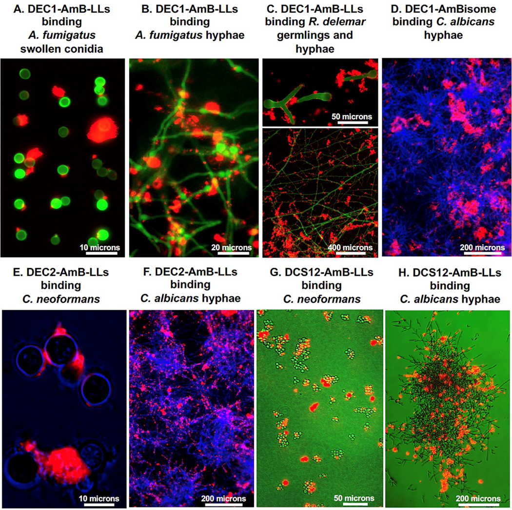
Fig. 4. Example images of DectiSomes binding to multiple developmental stages of diverse fungal pathogens.

(A & B). DEC1-AmB-LLs binding to A. fumigatus swollen conidia and hyphae, respectively. (C). DEC1-AmB-LLs binding to R. delemar germlings and mature hyphae. (D). DEC1-AmBisome binding to C. albicans hyphae. (E). DEC2-AmB-LLs binding to C. neoformans yeast cells. (F). DEC2-AmB-LLs binding to C. albicans hyphae. (G. & H). DC-SIGN isoform coated DCS12-AmB-LLs binding to C. neoformans yeast cells and C. albicans hyphae. All liposomes were tagged with Rhodamine B and the red epifluorescence of their binding is shown in red. A, B. GFP fluorescently labeled cells. C, D, F, G. Cell chitin is stained with calcofluor white and their fluorescence is shown in green or blue. E, H. Cells illuminated with bright field, but colored blue and green, respectively. C and G. Cells were grown and imaged of the surface of agar. These are replicate images of cell staining not shown in the original publications [107,215,217,218,219,220]. Size bars indicate the degree of magnification.
