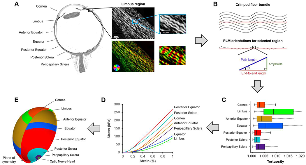
Figure 2. General strategy.

We divided the corneoscleral shell into seven regions (A). Each region was imaged using polarized light microscopy (PLM). The close-up images show the collagen fibers in the limbus region. The wavy pattern of collagen fibers is discernible and emphasized using a color map of orientation. To quantify collagen crimp, a straight line was marked manually along a fiber bundle (B). Trigonometric identities were then used pixel by pixel along the line to calculate local amplitude, path length, and end-to-end length based on the orientation information derived from PLM as described in detail in (Brazile et al., 2018; Gogola et al., 2018a; Jan et al., 2018). The local amplitude and lengths were then integrated to compute those of the fiber bundle. The crimp tortuosity of the fiber bundle was calculated as its integrated path length divided by the end-to-end length. We measured collagen crimp tortuosity in seven regions across the corneoscleral shell and the results (C) were fed into a fiber-based microstructural constitutive model to obtain region-specific nonlinear hyperelastic mechanical properties (D). A three-dimensional axisymmetric model (E) was then developed to simulate the IOP-induced deformation of the corneoscleral shell. The model-predicted tissue stretch was used to quantify collagen fiber recruitment over the corneoscleral shell.
