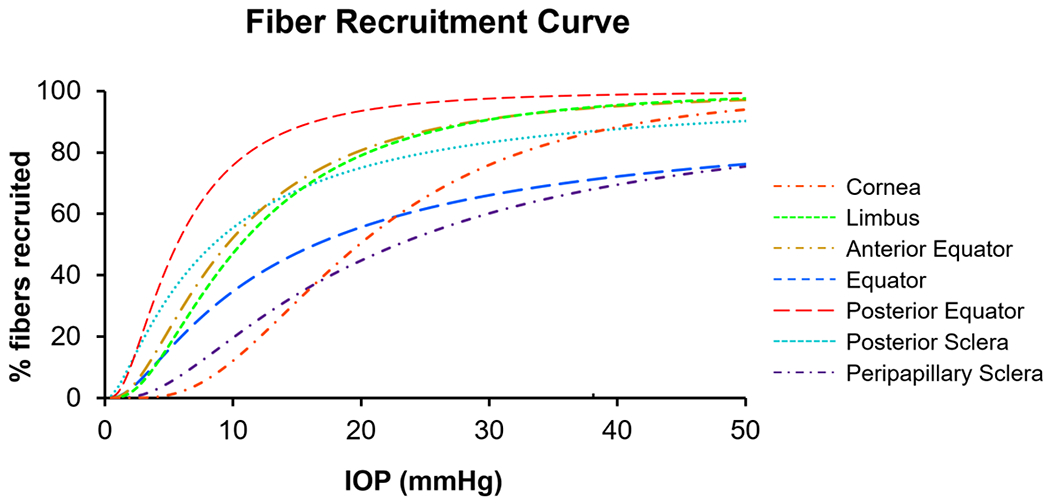
Figure 5.

The IOP-induced collagen fiber recruitment over the corneoscleral shell. The curves were colored by regions across the corneoscleral shell. All regions exhibited sigmoid recruitment curves, but the recruitment rates varied substantially. At low IOPs, collagen fibers in the posterior equator were recruited the fastest, such that at a physiologic IOP of 15 mmHg, over 90% of fibers were recruited, compared with only a third in the cornea and the peripapillary sclera. At an elevated IOP of 50 mmHg, collagen fibers in the limbus and the anterior/posterior equator were almost fully recruited, compared with 90% in the cornea and the posterior sclera, and 70% in the peripapillary sclera and the equator.
