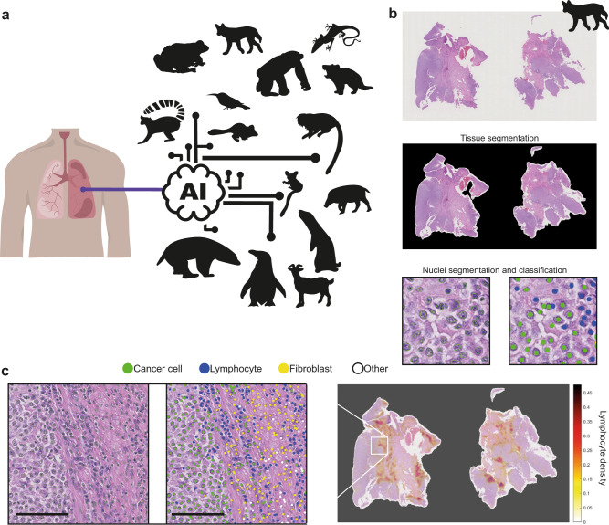
Fig. 1. Pan-species computational pathology.
a Transfer learning of cell identification from human lung to pan-species tumour pathology. b, c Overview of the H&E single-cell analysis pipeline as illustrated using a dog’s (CANFAM) transmissible venereal tumour sample. b This AI pipeline35 first segments the viable tissue area, then detects and classifies all cells into cancer, stromal, lymphocyte and others. For more details, see Methods. c The pipeline is implemented to spatially profile the immune microenvironment at the whole-slide level (right, immune cell density, cells/pixels2), after single-cell segmentation (left) and cell classification (middle). Scale bar, 50 µm. Cell colours are denoted as four classes, green: cancer (malignant epithelial) cells; blue: lymphocytes (including plasma cells); yellow: noninflammatory stromal cells (fibroblasts and endothelial cells); white: ‘other’ cell class that included nonidentifiable cells, less abundant cells such as macrophages and chondrocytes and ‘normal’ pneumocytes.

