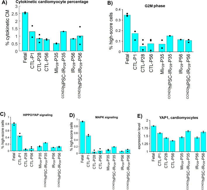
Figure 7.
Single-nuclei RNA sequencing shows that CCND2hiPSC-MI cardiomyocytes increase cycling and upregulated HIPPO/YAP & MAPK signaling pathways, especially 7 days after MI injury. (A) Percentage of cardiomyocytes highly expressing cytokinesis-specific genes AURKB, ALKBH4, ANLN, CNTROB, and KLHDC8B in each group. (B–D) Bar graphs: sparse analysis quantifies the G2M phase, HIPPO/YAP, and MAPK signaling pathways in each heart; here, the sparse model only used DNA synthesis genes to compute a 'sparse model score' for each cell such that the score optimally separates fetal from naïve-P56 cardiomyocytes; a higher score implies more active G2M, HIPPO/YAP and MAPK activities. Each dot is the percentage of cells having 'high model score' in a heart. (E) Error bar: YAP1 average expression, which was per cell in each group; here, the raw counts were logarithm (base 2) transformed and scaled according to the total of UMIs and detected genes per cell by Seurat30.

