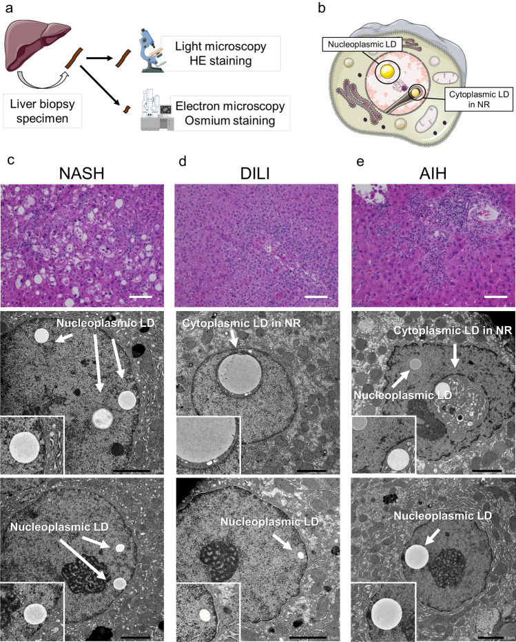
Figure 1.
Representative cases of nLDs and cLDs in NR. (a) Schematic depiction of the collection of liver biopsy samples and their processing for both hematoxylin and eosin (H&E) staining for light microscopy and osmium staining for electron microscopy. (b) Schematic illustration of nLDs and cLDs in NR. (c–e) Biopsy sections from patients with (c) NASH showing nLDs in hepatocytes and those from patients with (d) DILI and (e) AIH showing both nLDs and cLDs in NR. AIH autoimmune hepatitis, cLDs in NR cytoplasmic lipid droplets in the nucleoplasmic reticulum, DILI drug-induced liver injury, NASH nonalcoholic steatohepatitis, nLDs nucleoplasmic lipid droplets.

