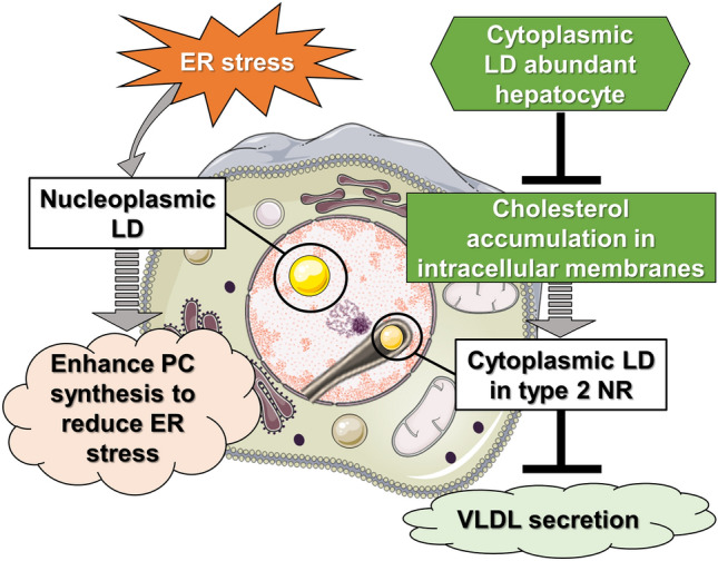
Figure 4.

Model depicting two distinct interpretations of nuclear LDs in liver diseases. Schematic representation of potential significance of LDs in the nucleus. Left model: in human hepatocytes under ER stress, secretion of VLDL can be suppressed, leading to accumulation of lipoprotein precursors in the ER lumen. These precursors then translocate into the lumen of type 1 NR (INM invagination), which results in increased amount of nLDs. The surface of nLDs can play a role in induction of PC synthesis to reduce ER stress in various liver diseases. Right model: alternatively, in hepatocytes of patients with low total and LDL cholesterol plasma levels, cholesterols will be sequestered in cellular membranes (e.g. in the NE), which may accelerate the formation type 2 NR (invagination of ONM and INM). cLDs may be initially packed in type 2 NR to be isolated from ER contact sites. This leads to decreased usage of lipids stored in cLDs for VLDL production in the ER. In these circumstances, cLDs in NR may negatively regulate the VLDL secretion machinery in hepatocytes. This machinery may be inhibited when patients’ liver show severe steatosis in which hepatocytes accumulate cholesterols as cholesterol esters in excessive cLDs instead of as free cholesterol in cellular membranes. cLDs in NR cytoplasmic lipid droplets in the nucleoplasmic reticulum, ER endoplasmic reticulum, INM inner nuclear membrane, LDL low-density lipoprotein, nLDs nucleoplasmic lipid droplets, NR nucleoplasmic reticulum, NE nuclear envelope, ONM outer nuclear membrane, PC phosphatydilcholine, VLDL very low-density lipoprotein.
