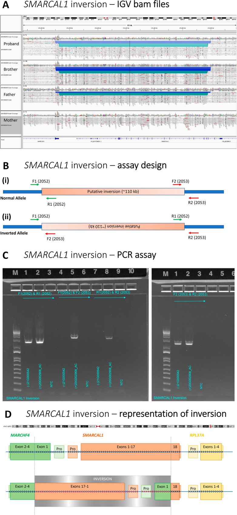
Fig. 3. Detection and confirmation of chromosome 2 inversion involving SMARCAL1.

A Integrative Genomics Viewer (IGV) WCR-free WGS read data visualisation showing the four family members and the site of the inversion; B Assay design for orthogonal confirmation demonstrating the inversion; C PCR assay demonstrating the presence or absence of inversion breakpoints in the family members; D Representative diagram demonstrating the inversion involving Exons 1–17 (of 18) of SMARCAL1 and the 5′UTR and beginning of exon 1 (p.1–79) of MARCHF4; Pro – Promoter region of corresponding gene; arrows indicate the standard 5′–3′ read strand of each gene, black depicts standard with red depicting the reversed direction of the gene after the structural variant, actual reading frame of the fusion gene may differ.
