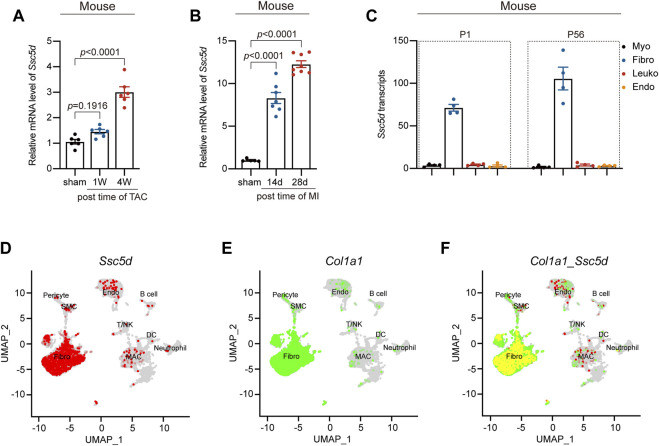
FIGURE 2.
Ssc5d mRNA levels were significantly elevated in MI and TAC mouse models. (A,B) The relative mRNA expression of Ssc5d after TAC (1 and 4 W) (n = 6) and MI (14 and 28 days) (n = 5–7) surgery. (C) Normalized Ssc5d expression (CPM) in each cell type at neonatal (P1) or adult stages (P56) mouse hearts (GSE95755). Myo (black), cardiomyocytes; Fibro (blue), fibroblasts; Leuko (red), leukocytes; Endo (orange), endothelial cells. (D–F) UMAP plot of Ssc5d and Col1a1 co-expression in sham mice cardiac non-cardiomyocyte clusters. Red, Ssc5d; Green, Col1a1; Yellow, Ssc5d and Col1a1 co-expression. Fibro, fibroblasts; SMC, smooth muscle cell; MAC, macrophage; DC, dendritic cell; Endo, endothelial; CPM, counts per million; W, week; d, day. p-value < 0.05 was considered statistically significant.

