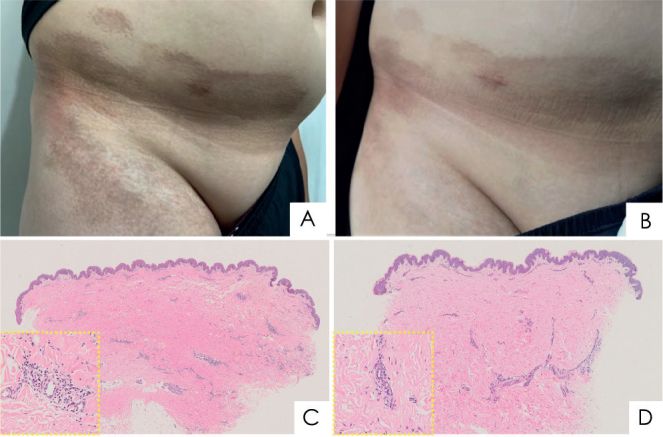
Fig. 2.

Clinical and histological features for case 2: (A, C) before and (B, D) after tofacitinib treatment for 5 months. (A) Before treatment, brown patches were observed on the right lower abdomen, groin and thigh. (B) After treatment, the skin lesions became softer and pigmentation considerably improved. (C) Histopathological examination of an abdominal skin biopsy revealed thickened and homogenous collagen bundles in the mid- and reticular dermis, with sparse deep lymphoplasmacytic infiltrate. Loss of adventitial fat resulted in a lack of eccrine glands. (D) Post-treatment biopsy specimens revealed decreased collagen bundle size and collagen fibres in the lower dermis (haematoxylin–eosin, original magnification ×4, enlarged view ×20 in yellow boxes of C and D).
