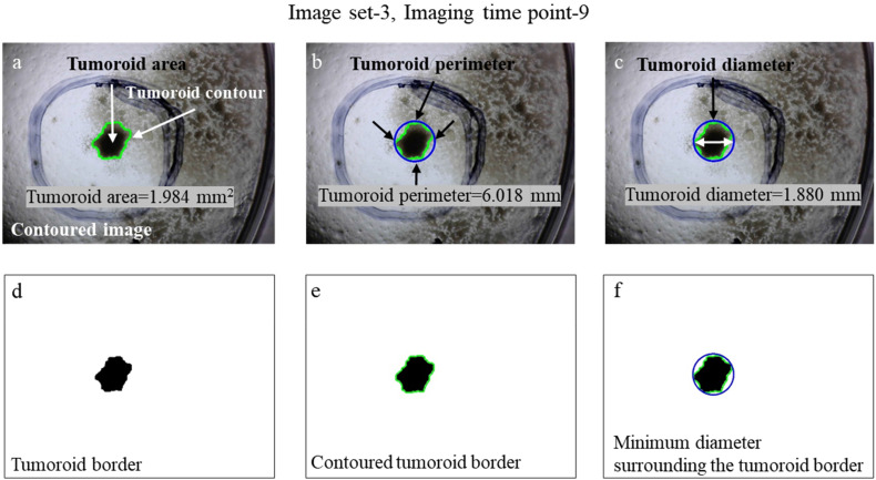
Figure 5.
Feature extractions of the 3 image sets, that is, Image set-3, Imaging time point-9. (a) Contouring and calculating the area of tumoroid, that is, tumoroid area of image set-3 at imaging time point-9 was 1.984 mm2. (b) The perimeter of tumoroid was 6.018 mm. (c) The diameter of tumoroid was 1.880 mm. (d–f) Binary formats of tumoroid border, contoured tumoroid border, and minimum diameter surrounding the tumoroid border.

