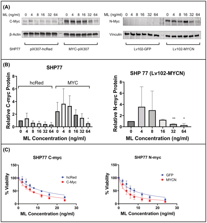
FIGURE 3.

Forced expression of myc sensitizes SCLC lines to ML. (A) Immunoblot of transduced SHP77 cells with C‐Myc and N‐myc following incubation with indicated concentrations of ML. (B) Graphic representation of C‐myc and N‐myc protein expression shown in panel A, calculated from three independent experiments. Relative C‐myc and N‐myc protein expression were normalized to loading control. SHP77 cells transduced with Lv102‐GFP do not have detectable N‐myc expression and are therefore not represented on the graph. *p < 0.05, **p < 0.01 for comparison with PBS group. (C) Cell viability of transduced SHP77 with the MTS assay following a 48‐h incubation with increasing concentrations of ML.
