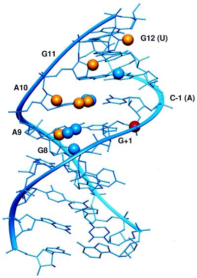
Figure 2.
NAIM compared with the stem A NMR structure. Sites of interference are denoted by spheres. Blue spheres represent sites of interference consistent with the structure. The yellow spheres represent sites that cannot be interpreted in comparison with the structure. The scissile phosphate is denoted by a red sphere.

