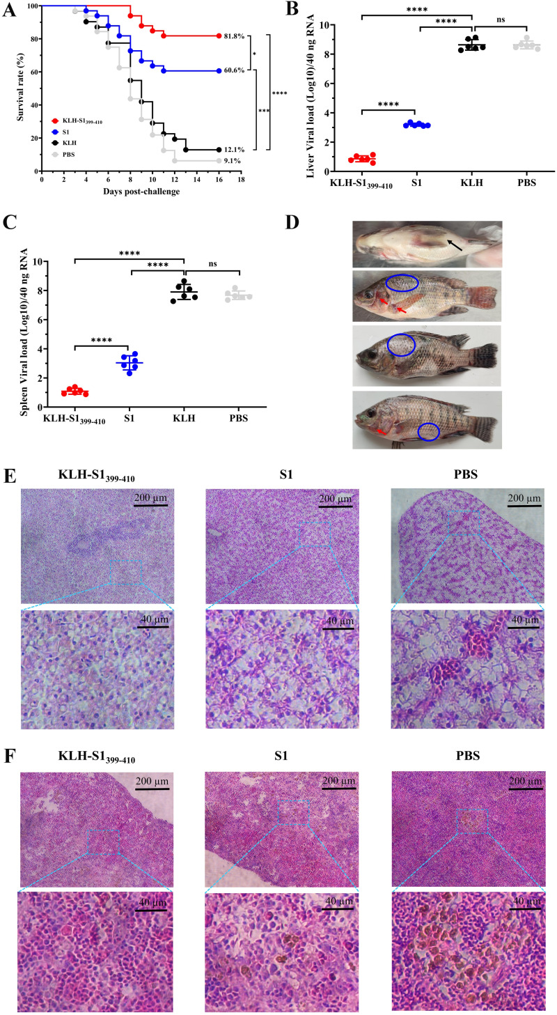
FIG 5.
Epitope-based synthetic peptide vaccine acted efficiently as prophylactic against TiLV infection. (A) Survival rate of different immunization groups after 15 days of intraperitoneal challenge with a lethal dose of TiLV. The survival curve was calculated by GraphPad Prism software. (B and C) Viral load of TiLV in liver (B) and spleen (C) tissues were quantified by qRT-PCR at 8 days after challenge. Data are means ± SD (n = 6 per group). (D) Representative figures showing typical symptoms of TiLV infection in tilapia, including abdominal swelling (black arrows), hyperemia (red arrows), and scales dropping off (blue circle). (E and F) Histopathological changes of different immune groups after 8 days of challenge were observed by H&E staining. Hepatocytes (E) showing fibrosis with polarized nuclei and aggregation were observed in the S1-immune group (middle), whereas the PBS group (right) showed severe hepatocyte fibrosis, nuclear polarization, and syncytial liver symptoms, and with hepatic congestion. Spleen tissue (F) showed few (S1 immunization group) or large aggregates (PBS group) of the melanomacrophage center (MMC) and vacuolated nucleus. Statistical significance was tested by one-way ANOVA and Tukey’s multiple-comparison tests. ****, P < 0.0001; ***, P < 0.001; **, P < 0.01; *, P < 0.05.

