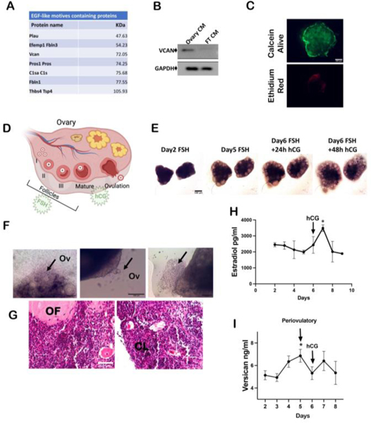
Figure 4: The EGF-like domain containing proteoglycan versican is secreted during ovulation in a 3D dynamic culture.
(A) Table showing proteins between 50–100 kDa containing EGF-like domains that were identified in ovarian conditioned media through mass spectrometry. (B) Western blot analysis of versican expression in medium conditioned with ovary and fallopian tube (FT) (CM=conditioned media). (C) Live/Dead staining using permeable calcein dye (green) and ethidium red (red). (D) Schematic of ovulation induced by FSH/hCG stimulation. (E) Bright field images of ovarian follicles growth during the treatment for 6 days treatment with FSH. (F) Bright field images of ovulating follicles. Black arrow pointing at oocytes extrusion. (G) Hematoxylin & Eosin staining showing follicles maturation. Ov=ovulation; OF= ovulated follicle; CL= corpus luteum. (H) ELISA detecting estradiol levels in conditioned media collected during the ex vivo dynamic ovulation on PREDICT. (I) ELISA detecting versican levels in conditioned media collected during the ex vivo dynamic ovulation on PREDICT. A minimum of three independent experiments, biological replicates were analyzed using one-way ANOVA (p*<0.05).

