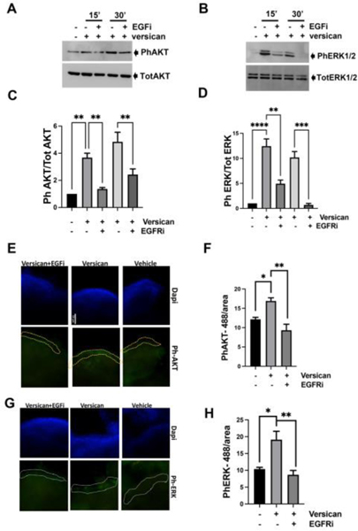
Figure 7: Versican activates EGFR signaling in fallopian tube epithelium cells.
Western blot analysis of phosphorylated AKT and phosphorylated ERK1/2 using specific antibodies in MOE cells (A-C) and FTE 33-Tag cells (B-D) incubated with versican 100 ng/ml for 15 and 30 minutes −/+ EGFR inhibitor 1 μM. Immunofluorescence analysis of phosphorylated AKT (E-F) and phosphorylated ERK1/2 (G-H) using specific antibodies in murine oviductal organs cultured in the PREDICT microfluidic device. A minimum of three independent experiments, biological replicates were analyzed using one-way ANOVA (ns>0.05, p*<0.05, p**<0.01, p***<0.001, P****<0.0001).

