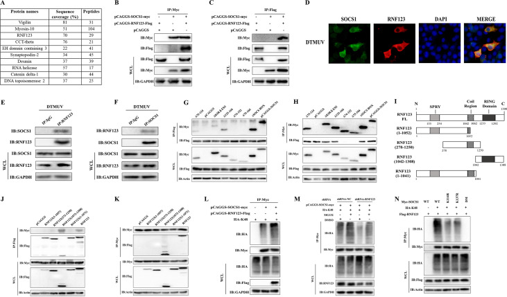
FIG 2.
RNF123 binds to the SH2 domain of SOCS1 through its RING domain and promotes the K48-linked ubiquitination of SOCS1. (A) Mass spectroscopic analysis of potential SOCS1 binding proteins. The numbers of peptides recognized in interacting proteins (including RNF123) are shown. (B and C) HEK 293T cells were transfected with pCAGGS-RNF123-Flag (1.5 mg/well) and pCAGGS (1.5 mg/well), pCAGGS and pCAGGS-SOCS1-Myc (1.5 mg/well), and pCAGGS-RNF123-Flag and pCAGGS-SOCS1-Myc. Thirty-six hours later, the cell lysates were immunoprecipitated with anti-Myc or anti-Flag, followed by IB with anti-Myc and anti-Flag. (D) When the cell confluence reached about 90%, the cells were infected with DTMUV at an MOI of 1. Thirty-six hours after infection, the cells were collected for indirect immunofluorescence detection of endogenous SOCS1 and RNF123 colocalization (original magnification, ×600). (E and F) DEFs were infected with DTMUV at an MOI of 1. Thirty-six hours later, the cell lysates were immunoprecipitated with anti-SOCS1, anti-RNF123, or IgG, followed by IB with anti-SOCS1 and anti-RNF123. (G and H) HEK 293T cells were transfected with pCAGGS-RNF123-Flag (1.5 mg/well) together with the plasmids for full-length or truncated SOCS1 (1.5 mg/well). Thirty-six hours later, the cell lysates were immunoprecipitated with anti-Myc or anti-Flag, followed by IB with anti-Myc and anti-Flag. (I) Schematic representation of plasmids for full-length (FL) and truncated RNF123. (J and K) HEK 293T cells were transfected with pCAGGS-SOCS1-Myc (1.5 mg/well) together with the plasmids for full-length or truncated RNF123 (1.5 mg/well). Thirty-six hours later, the cell lysates were immunoprecipitated with anti-Myc or anti-Flag, followed by IB with anti-Myc and anti-Flag. (L) HEK 293T cells were transfected with HA-Ub-K48 (1 mg/well) together with pCAGGS-SOCS1-Myc (1 mg/well) or pCAGGS-RNF123-Flag (1 mg/well) and pCAGGS-SOCS1-Myc. Thirty-six hours after transfection, the cell lysates were immunoprecipitated with anti-Myc, and ubiquitinated SOCS1 was detected by IB with anti-HA. (M) HEK 293T cells were transfected with pCAGGS-SOCS1-Myc (1 mg/well) and HA-Ub-K48 (1 mg/well) together with shRNA-RNF123 (1 mg/well) or shRNA-NC (blank control of shRNA-RNF123). Thirty-six hours after transfection, cells were treated with MG132 (10 μM) or DMSO for 6 h. The cell lysates were immunoprecipitated with anti-Myc, and ubiquitinated SOCS1 was detected by IB with anti-HA. (N) HEK 293T cells were transfected with pCAGGS-RNF123-Flag (1 mg/well) and HA-Ub-K48 (1 mg/well) together with the WT, the K114R or K137R single-point mutant, or the DM (1 mg/well). Thirty-six hours after transfection, the cell lysates were immunoprecipitated with anti-Myc, and ubiquitinated SOCS1 was detected by IB with anti-HA. All data are the mean values obtained from at least three replicates of independent experiments.

