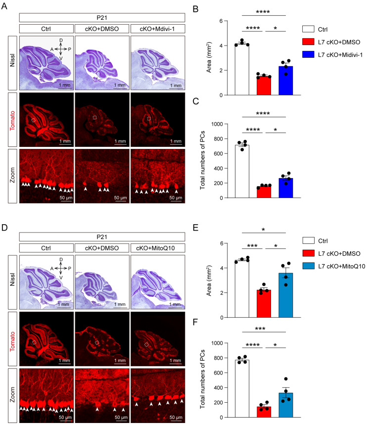
Figure 6.
Suppression of ROS production significantly rescue cerebellar PC loss in cKO mice. (A) Nissl staining of sagittal histological sections of the cerebellar vermis and immunofluorescent image of Tomato (red) staining to reveal PCs in the cerebellar vermis of the Ctrl and cKO mice treated with DMSO or the cKO mice treated with Midiv-10 (scale bar: 1 mm, 50 μm). (B) Quantification of the area of sagittal sections of the cerebellar vermis from the Ctrl and cKO mice (mean ± SEM; **** p < 0.0001, * p < 0.05, n = 4). (C) The number of PCs per sagittal histological section of the cerebellar vermis from the Ctrl and cKO mice (mean ± SEM; **** p < 0.0001, * p < 0.05, n = 4). (D) Nissl staining of sagittal histological sections of the cerebellar vermis and immunofluorescent image of Tomato (red) staining to reveal PCs in the cerebellar vermis of the Ctrl and cKO mice treated with DMSO or the cKO mice treated with MitoQ10 (scale bar: 1 mm, 50 μm). (E) Quantification of the area of sagittal sections of the cerebellar vermis of the Ctrl and cKO mice (mean ± SEM; *** p < 0.001, * p < 0.05, n = 4). (F) The number of PCs per sagittal histological sections of the cerebellar vermis of the Ctrl and cKO mice. (mean ± SEM; **** p < 0.0001, *** p < 0.001, * p < 0.05, n = 4). a, anterior; d, dorsal; p, posterior; v, ventral.

