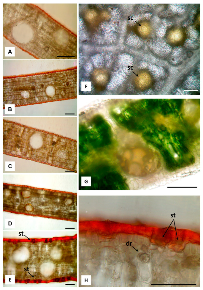
Figure 3.
Light microscopy images. (A–E) Hand-made transverse leaf sections stained with Sudan III highlight the thick layers of cuticle and the secretory cavities spread in the mesophyll: E. alba (A), E. eugenioides (B), E. fasciculosa (C), E. robusta (D), E. stoatei (E). (F,G) Leaves of E. alba: abaxial surface after bleaching with sodium hypochlorite, showing bright yellow secretory cavities (sc), and their overlying cells (arrows) (F); transverse section showing a secretory cavity containing many drops of brownish yellow EO (G). (H) Transverse section of E. stoatei leaf stained with Sudan III, showing a thick cuticle, and stomata (st); a druse (dr) under the epidermis is also visible. 100 µm bars.

