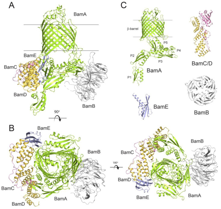
Figure 2.
Structures of BAM and the individual Bam proteins from E. coli. (A) The structure of fully assembled BAM (PDB ID: 5D0O) shown from the side, transmembrane view; membrane boundaries are indicated by the black lines. (B) Orthogonal views of BAM showing the extracellular, top view (left) and the periplasmic, bottom view (right). (C) The individual components BamA from N. gonorrhoeae (PDB ID: 4K3B) showing the β-barrel domain and the POTRA domains (P1-P5), BamB from E. coli (PDB ID: 3Q7N), the BamC/D complex from E. coli (PDB ID: 3TGO), and BamE from N. gonorrhoeae (PDB ID: 5WAM).

