Figure 1.
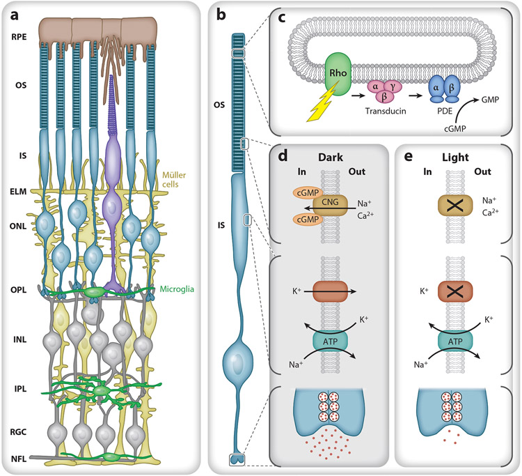
Retinal morphology and basic photoreceptor physiology. (a) Schematic of the retina from the outer (top) to inner (bottom) in cross section. The retinal pigment epithelium (RPE) (brown) lies between the vascular choriocapillaris and photoreceptor outer segments (OS). Müller cells (yellow) span the retina from the nerve fiber layer (NFL) to the photoreceptor inner segments (IS) and form the external limiting membrane (ELM) that helps to establish the subretinal space as a distinct microenvironment. Normally, microglia (green) reside in the outer plexiform layer (OPL), inner plexiform layer (IPL), retinal ganglion cell layer (RGC), and NFL. (b) Rod photoreceptor compartment. From top to bottom: OS containing stacks of discs, IS with metabolic and biosynthetic machinery, cell body, and synaptic terminal. (c) A photon causes rhodopsin to change conformation, activating transducin (Gαtβ1γ1). Each GTP-bound Gαt then binds and activates cGMP phosphodiesterase 6 (PDE), allowing it to hydrolyze cGMP, decreasing intracellular cGMP levels. (d) In the dark, cGMP opens a cyclic nucleotide gated (CNG) channel in the OS plasma membrane, allowing cationic influx (Na+, Ca2+) that is balanced by an efflux of cations, mostly potassium (K+), from the IS. The IS Na+/K+ transporter uses ATP to maintain the electrochemical gradient and depolarized membrane potential, leading to the continual release of glutamate. (e) In light, cGMP levels fall, and the CNG channels close, reducing the influx of Na+ and Ca2+ and causing the cell to hyperpolarize and reduce glutamate release. Additional abbreviations: INL, inner nuclear layer; ONL, outer nuclear layer.

