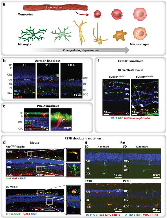
Figure 2.
Variations in the response of microglia and infiltrating monocytes during photoreceptor degeneration. (a) Microglia are typically ramified in healthy tissue, with small cell bodies and numerous thin processes (green). Activated microglia lose their branched morphology and migrate (yellow) to the afflicted region of damage, often transitioning into a macrophage phenotype (tangerine). Infiltrating bone marrow–derived monocytes (red) rapidly differentiate into macrophages (pink), becoming difficult to distinguish from resident immune cells. (b) In Arrestin-1 knockout mice, light onset causes microglia and monocytes (green, green + red) to migrate to the ONL and phagocytose photoreceptor cells bodies (96 h), eliminating the ONL within a week and leaving a handful of subretinal macrophages between the RPE and the ELM (240 h). Iba1 (green) labels microglia and macrophages; CD11b (red) labels microglia and leukocytes (e.g., monocytes); and nuclei are stained with DAPI (blue). Panel adapted from Karlen et al. (2018) (CC BY-SA 4.0). (c) In PRCD knockout mice, microglia (red) primarily target the inner (unlabeled)–outer (green) segment junction, with their processes extending toward the cilium (green). P2YR12 (red) labels microglia; CD68 (purple) labels lysosomes; WGA (green) labels the OS; and nuclei are stained with Hoescht (blue). Panel adapted with permission from Spencer et al. (2019). (d) In P23H rhodopsin mice (top) and LD mice (bottom), microglia with upregulated Gal3 (green + red) accumulate in the subretinal space, whereas infiltrating monocytes (green) remain in the retina. Iba1 (top, green) labels microglia and macrophages; YFP (Cx3CR1) (bottom, green) labels Cx3CR1+ cells (e.g., microglia, macrophages); GAL3 (red) labels subretinal macrophages; and nuclei are stained with DAPI (blue). Panel adapted with permission from O’Koren et al. (2019). (e) In P23H rhodopsin rats, microglia (green, green + red) remain in the subretinal space throughout adulthood, indicating chronic neuroinflammation after photoreceptor degeneration has been completed; compare 4 months (left) to 12 months (right) in controls (SD, top) and P23H rhodopsin mutants (bottom). Iba1 (green) labels microglia and macrophages; MHC-II RT1B (red) label macrophages; and nuclei are stained with TO-PRO-3 iodide (blue). Panel adapted from Noailles et al. (2016) (CC BY-SA 4.0). (f) In aged, 18-month Cx3CR1 knockout mice exposed to normal 12 h on/off light levels, microglia (green) appear in the inner retina (both conditions), and additional microglia accumulate in the subretinal space of the knockout (Cx3CR1GFP/GFP) (arrows). GFP (green) indicates Cx3CR1-positive cells; Griffonia simplicifolia-positive (red) labels vascular endothelial cells; and nuclei are stained with DAPI (blue). Panel adapted with permission from Combadière et al. (2007). Abbreviations: ELM, external limiting membrane; GFP, green fluorescent protein; INL, inner nuclear layer; IPL, inner plexiform layer; IS, inner segment; LD, light damage; ONL, outer nuclear layer; OPL, outer plexiform layer; OS, outer segment; PRCD, progressive rod–cone degeneration; RGC, reginal ganglion cell; RPE, retinal pigment epithelium; SD, Sprague-Dawley control rats; WGA, wheat germ agglutinin.

