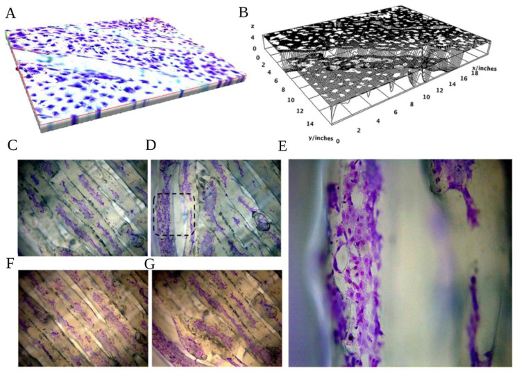
Figure 6.
(A) Spatial representation of the poly(ε-caprolactone) (PCL) compounded with beta-tricalcium phosphate 20% (PCL+β-TCP 20%)-based biomaterial with toluidine blue-stained preosteoblasts, magnification 20×. (B) Surface plot of the PCL+β-TCP 20% compounds with the relevant expansion of white dots (pre-osteoblasts) on almost the whole surface. (C) Toluidine blue staining of preosteoblasts expanding on the central meshes or the peripheral (D,E) meshes of the compound, magnification 20× (C,D) and 40× (E, as magnification of black dashed square of D). The hematoxylin/eosin staining of the proliferating preosteoblasts on the biomaterial network better elucidates the affinity of the compound with the developing cells in the centre (F) or peripheral parts of the substrate (G).

