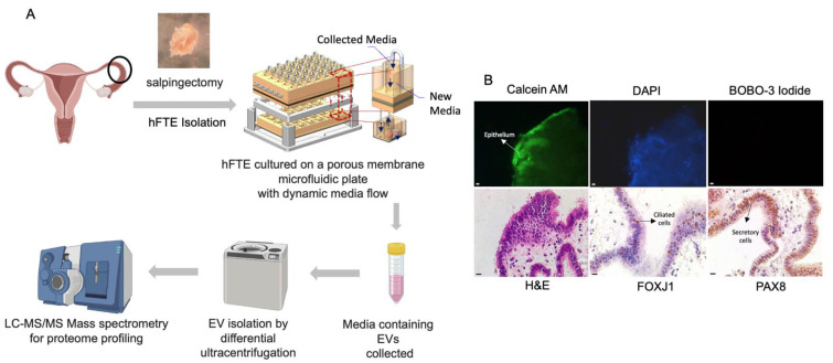
Figure 1.
The fallopian tube explants were cultured in a microfluidic platform for sEV collection. (A) The workflow of hFTE-derived EV proteomics characterization. (B) The live/dead staining of hFTE epithelium cultured in the PREDICT-MOS for six days. The green fluorescence (calcein) indicates live cells, and the red fluorescence indicates dead cells, labeled by BOBO-3 iodide. Immunohistochemistry staining of FOXJ1 and PAX8 confirmed that the hFTE maintained the two major epithelial cell populations, ciliated and secretory cells, after six-day dynamic culture on the PREDICT-MOS. Scale bar = 10 µm.

