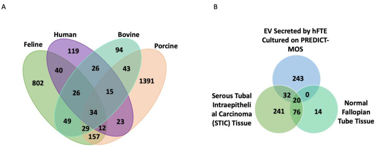
Figure 4.
hFTE sEV protein content comparison across species and FT lesions. (A) Venn diagram showing the number of sEV proteins identified exclusively in human fallopian tube epithelium (hFTE), bovine, porcine, cat oviduct, or in common [3,33,34]. (B) Venn diagram showing shared proteins identified in STIC tissue [35] and proteins found in hFTE-derived sEVs cultured in the microfluidic device, PREDICT-MOS.

