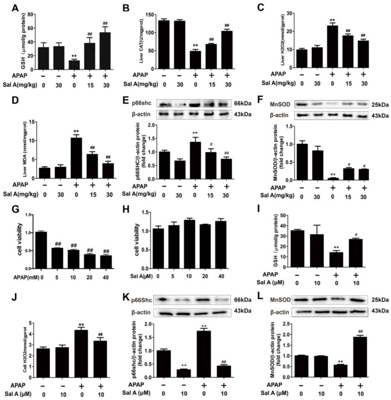
Figure 2.
Sal A ameliorates hepatic oxidative stress in vivo and in vitro. (A–F) Sal A pretreatment was performed for 3 days followed by APAP stimulation in mice. (A–D) GSH, CAT, H2O2 and MDA levels in mice, n = 8. (E,F) p66Shc and MnSOD protein expression in mice, n = 3. (G) AML12 cells were exposed to different concentrations of APAP (0, 5, 10, 20 and 40 mM) for 24 h. Cell viability, n = 8. (H) AML12 cells were exposed to Sal A for 24 h at different concentrations of 0, 5, 10, 20 and 40 µM. Cell viability, n = 8. (I–L) AML12 cells were incubated with Sal A before exposure to APAP. (I,J) GSH and H2O2 levels in AML12 cells, n = 8. (K,L) p66Shc and MnSOD protein expression in AML12 cells, n = 3. ** p < 0.01 vs. control. # p < 0.05, ## p < 0.01 vs. APAP.

