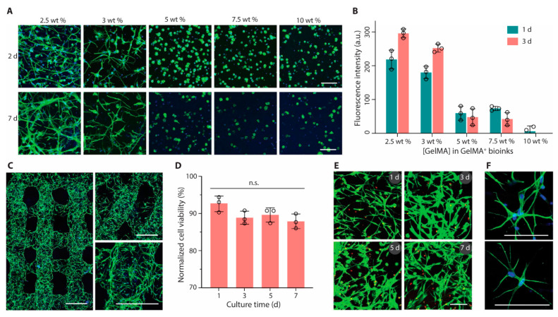
Figure 2.
Bioprinting and culture of constructs containing astrocytes. (A) Fluorescence microscopy images (green, phalloidin; blue, DAPI) and (B) metabolic activity of astrocytes encapsulated in GelMA+ hydrogels with varied GelMA concentrations (2.5 to 10 wt%). (C) Confocal laser scanning microscopy images (green, phalloidin; blue, DAPI) of printed multilayer lattices with 90° or 60° angles between adjacent layers (2 days). (D) Cell viability and (E) Live/Dead-stained astrocytes (green, calcein AM; red, ethidium homodimer-1) after bioprinting and culture for up to 7 days. (F) High-magnification fluorescence microscopy images of astrocytes (green, phalloidin; blue, DAPI) in printed constructs (2 days). GelMA+ (2.5 wt%) and astrocytes were used for bioprinting in (C) to (F). Scale bars, 100 μm (A,E,F) and 500 μm (C). Reprinted with permission from ref [73].

