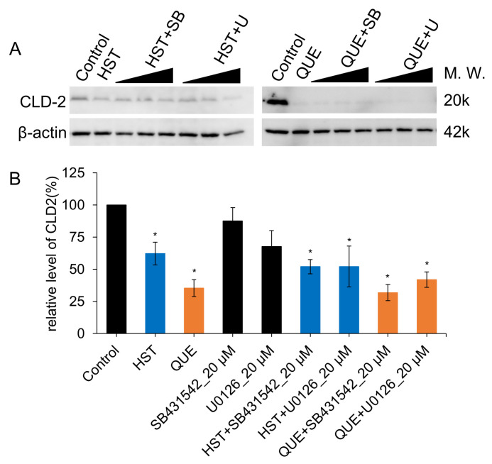
Figure 7.
Changes in the relative amount of protein of CLD-2 after compounds treatment for 48 h in MDCK II cells. (A) Western blotting analysis of CLD-2 expressed in control (DMSO) and 100 µM flavonoid- and/or 5, 10, 20 µM SB431542/U0126 -treated MDCK II cells. (B) Quantitative analysis with densitometry of 100 µM flavonoid- and/or 5, 10, 20 µM SB431542/U0126- treated MDCK II cells. Turkey-Kramer multiple comparison tests were applied as statistical analyses. Error bars show standard deviations. Different from the value of the control cells, * p < 0.05. HST, QUE: n = 8.

