Figure 3.
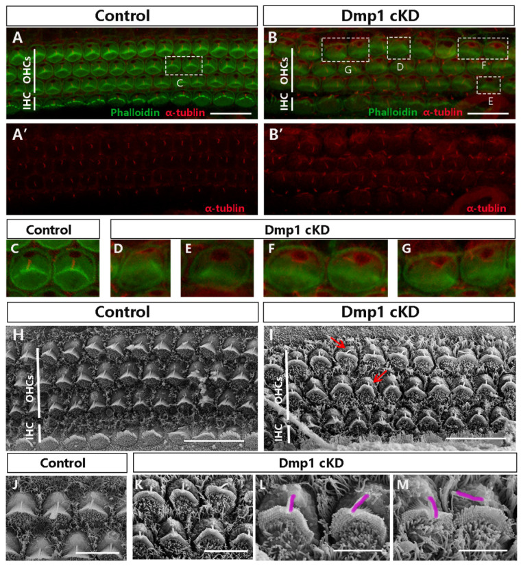
The proper positioning of kinocilia was affected in Dmp1 cKD mice. (A,B): Confocal microscope images from the basal regions of Dmp1 cKD and littermate control mice cochlea, Acetylated tubulin staining was singly showed in (A′,B′) (Acetylated tubulin: red. Phallodin: green.) (C–G): The amplification of the white dashed box in (A,B). In normal control cochlea, hair cells had a single kinocilium located at the vertex of the stereociliary bundles (C). By contrast, in the Dmp1 cKD mice, the kinocilia were misplaced and deviated from the vertex of the V-shape hair bundles, and the bundles were deformed, with no clear vertex (D,F,G). Several hair cells also had deletions of kinocilium (E). (H–M): SEM of outer hair cells from basal cochlear turns of Dmp1 cKD and control mice at P1 using scans with different magnifications. (H,I): 3 k magnification. (J,K): 8 k magnification. (L,M): 15 k magnification. Red arrows showed the abnormal positioning of kinocilia.Under the higher magnification, the separation between kinocilia and stereociliary bundles was clearly noted. The kinocilia were outlined (purple). Scale bars: (A–I): 10 µm; (J,K): 5 µm; and (L,M): 3 µm.

