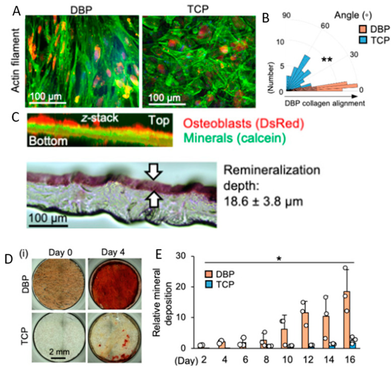
Figure 4.
Osteoblasts grown on demineralized bone paper (DBP) display enhanced regenerative capacities compared to osteoblasts grown on tissue culture polystyrene (TCP). (A) Morphology of OBs grown on vertically sectioned DBP and TCP for one week. (B) Immunofluorescent staining of actin filaments. (C) Circular histogram of cell alignment angles (n = 100). (C) Top: z-staked cross-sectional image of DBP will osteoblasts growing. Bottom: Cross section of 100-μm-thick DBP stained with alizarin red after three-week culture of osteoblasts (n = 3). (D) Mineral deposition by osteoblasts on DBP and TCP, showing alizarin red mineral stain on days zero and four. (E) Time-course measurement of mineral deposition by osteoblasts for 16 days (n = 3). * indicates p-value < 0.05; ** indicates p-value < 0.01. Figure adapted with permission under a Creative Commons Open Access License from Park et al., 2021 [120].

