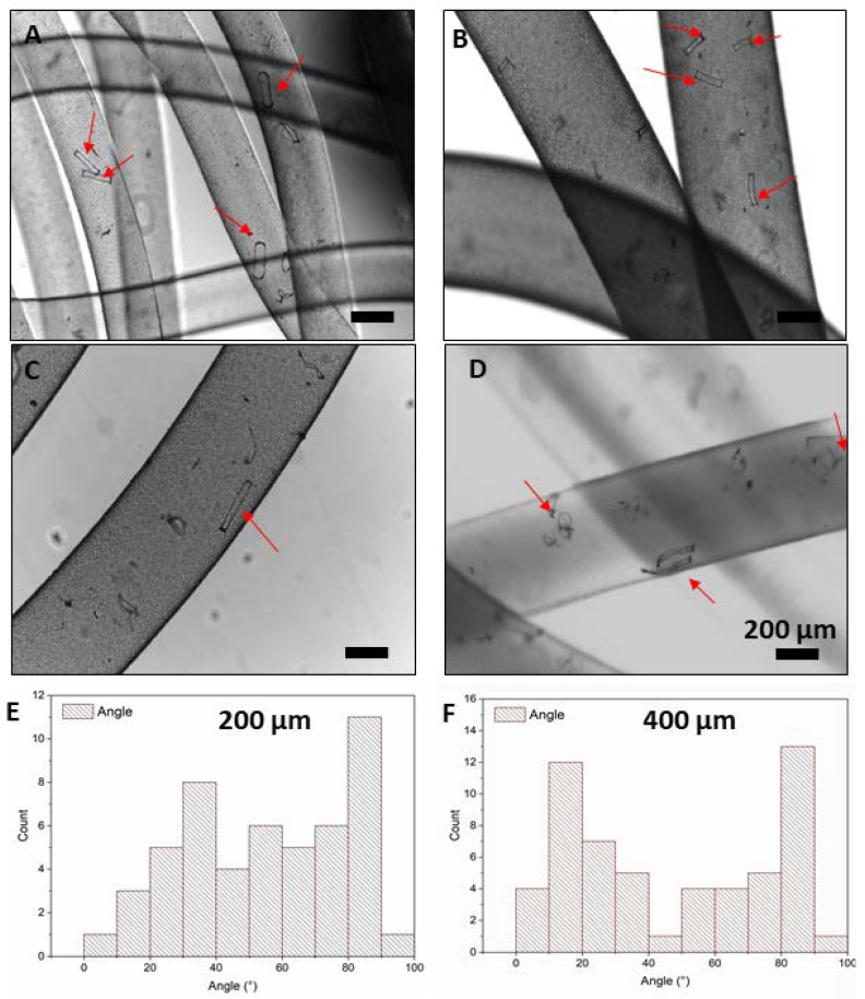
Figure 5.
Images taken by optical microscopy show the morphology of the hydrogel-based filaments (ADA-GelMA, 2:1, 10 wt%) with rod-shaped fillers of (A,B) 200 and (C,D) 400 μm length. These pictures are taken from different spots of the spun filaments. The red arrows spot the rods within the wet spun filaments. Angular distribution for the (E) 200 μm and (F) 400 µm length microrods (14,000 rods/mL) with respect to the fiber axis.

