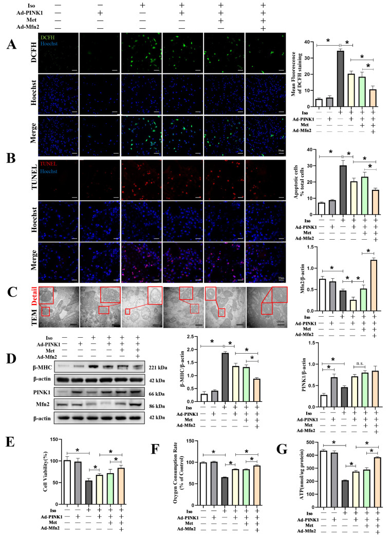
Figure 6.
Mfn2 and Metformin in collaboration with PINK1 improved mitochondrial function and prevented Iso-induced cardiomyocyte injury. Cardiomyocytes were split into six groups: (1) control + Ad-control, cardiomyocytes were transfected with control adenovirus and were not treated with Iso; (2) control + Ad-PINK1, cardiomyocytes were treated with PINK1 adenovirus and were not treated with Iso; (3) Iso + Ad-control, cardiomyocytes were transfected with control adenovirus and treated with 10 µM Iso for 48 h; (4) Iso + Ad-PINK1, cardiomyocytes were transfected with PINK1 adenovirus and treated with 10 µM Iso for 48 h; (5) Iso + Ad-PINK1 + Met, cardiomyocytes were transfected with PINK1 adenovirus and treated with 10 µM Iso and 2 mM metformin for 48 h; and (6) Iso + Ad-PINK1 + Met + Ad-Mfn2, cardiomyocytes were transfected with PINK1 and Mfn2 adenovirus then treated with 10 µM Iso and 2 mM metformin for 48 h. (A) DCFH staining was used to show ROS production in cardiomyocytes. DCFH-positive cells are shown in green. (B) TUNEL staining was used to show apoptotic cardiomyocytes in each group. TUNEL-positive cells are shown in red. (C) Mitochondrial morphology is shown by TEM in cardiomyocytes. Details of mitochondrial structures are shown in the red frame. (D) Western blot showing the expression of β-MHC, PINK1 and Mfn2 in cardiomyocytes. (E) CCK-8 assay results show the cell viability in cardiomyocytes. (F) Mitochondrial respiration was measured using extracellular oxygen consumption assay kits by assessing oxygen consumption rate. (G) ATP assay kit with a luminometer was used to determine intracellular ATP levels. Statistical analyses (A–G) were performed by one-way ANOVAs followed by Bonferroni test post-hoc tests. Data are presented as the means ± SD, (n = 3). * p < 0.05.

