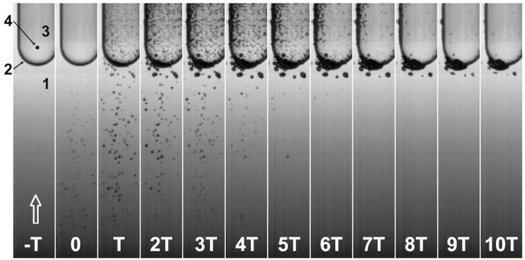
Figure 1.
Sequence of high-speed images (T ≈ 33.3 µs) showing cavitation bubbles inside a sealed vial containing ethanol after exposure to a single-pulse shock wave with a positive peak amplitude of approximately 66 MPa. The arrow shows the direction of the shock wave, which propagated through water (1), penetrated the vial at its bottom (2), and continued its path through the ethanol (3). The focus F was located at the center of curvature of the vial (4). The first frame where bubbles could be observed was t = 0.

