
Figure 3.
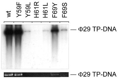
Φ29 DNA polymerase mutants are strongly affected in Φ29 TP–DNA amplification. The assay was carried out as described in Materials and Methods, in the presence of 5 ng wild-type or mutant Φ29 DNA polymerase, 5 ng Φ29 TP and 10 µg each Φ29 DBP and Φ29 SSB. After incubation for 90 min at 30°C, samples were processed and the amplified DNA was analyzed by alkaline agarose gel electrophoresis as described in Materials and Methods. (Top) Autoradiograph of such a gel; (bottom) Φ29 TP–DNA amplification signal detected by ethidium bromide staining. The migration position of unit length Φ29 TP–DNA (19 285 bases) is indicated.