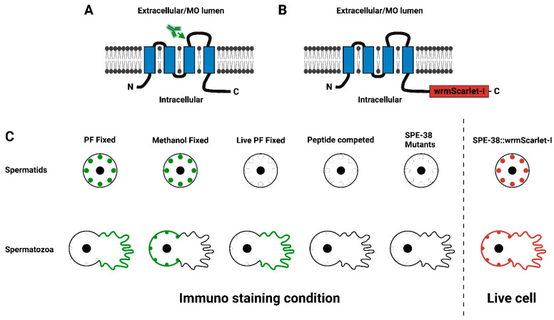
Figure 1.
(A,B) A schematic representation of the C. elegans SPE-38. The four transmembrane domains are indicated by the blue boxes imbedded in the membrane. The N-Terminus and C-Terminus of SPE-38 are intracellular while the loops between the first and second transmembrane domain and between the third and fourth transmembrane domain are extracellular or in the MO lumen. The antibody symbol and arrow (A) indicate the location of the peptide sequence used to raise the polyclonal antisera used by Chatterjee et al., 2005. (B) A schematic representation of SPE-38::wrmScarlet-I. The red box (not to scale) indicates the location of wrmScarlet-I. The wrmScarlet-I sequence is codon optimized for expression in C. elegans along with key sequence changes (-I isoleucine substitution) that improve molecule stability. (C) The left panel is a schematic summary of previous immunolocalization experiments and controls for antisera specificity published in Chatterjee et al., 2005. Green indicates antibody staining in spermatids and mature sperm. In the right panel red indicates the observed distribution of SPE-38::wrmScarlet-I in spermatids and mature sperm.

