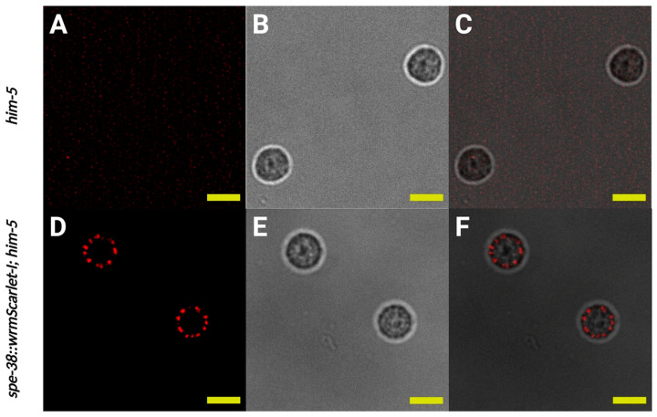
Figure 3.
Localization of SPE-38::wrmScarlet-I visualized by red fluorescent signal to membranous organelles (MOs) in spermatids. (A–C) Imaging of the same control spermatids dissected from him-5(e1490) males. (A) Single confocal section of control spermatids. (B) Bright field image of control spermatids. (C) Merge of (A,B). (D–F) Imaging of spe-38::wrmScarlet-I; him-5(e1490) spermatids dissected from males. (D) Single confocal section of spe-38::wrmScarlet-I; him-5(e1490) spermatids. (E) Bright field image of spe-38::wrmScarlet-I; him-5(e1490) spermatids. (F) Merge of (D,E). Yellow scale bars, 5 µm.

