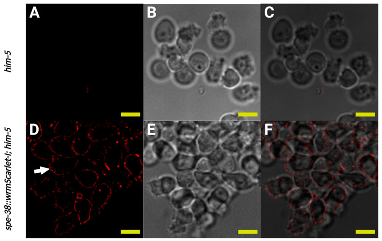
Figure 4.
SPE-38::wrmScarlet-I visualized by red fluorescent signal localizes to fused MOs, the cell body and pseudopod plasma membranes in spermatozoa. (A–C) Imaging of the same in vivo activated control spermatozoa dissected from mated hermaphrodites. (A) Single confocal section of control spermatozoa. (B) Bright field image of control spermatozoa. (C) Merge of (A,B). (D–F) Imaging of spe-38::wrmScarlet-I; him-5(e1490) in vivo activated spermatozoa dissected from mated hermaphrodites. (D) Single confocal section of spe-38::wrmScarlet-I; him-5(e1490) spermatozoa. White arrow indicates fused MOs in a spermatozoa. (E) Bright field image of spe-38::wrmScarlet-I; him-5(e1490) spermatozoa. (F) Merge of (D,E). Yellow scale bars, 5 µm.

