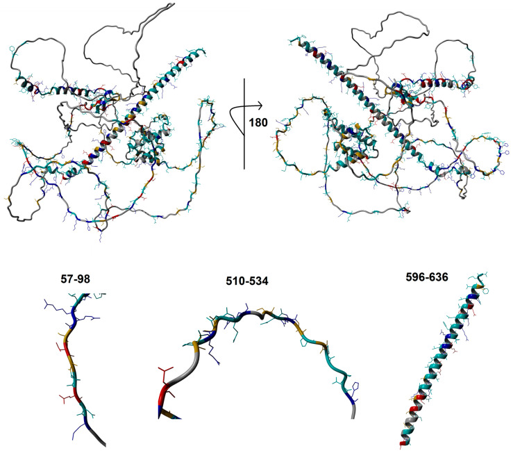
Figure 2.
Predicted WAC protein structure. An AlphaFold protein model of WAC showing conserved amino acids colored as blue: polar basic; red: polar acidic; orange: S/T; cyan: all other conserved amino acids. Shown below are zoomed in view of several conserved motifs within WAC that fall within intrinsically disordered regions.

