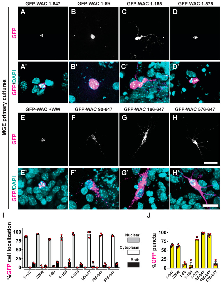
Figure 6.
Distinct cellular distribution by WAC’s conserved protein domains. MGE primary neurons transfected with GFP-WAC fusion proteins were assessed for GFP localization (A–H) and merged with DAPI after five days in vitro (A’–H’). (I) Quantification of the proportion of GFP labeled cells showing nuclear and/or cytoplasmic distribution. (J) Quantification of the proportion of GFP-labeled cells with punctate localization. Scale bars in (H) = 40 µm for all top panels and (H’) = 20 µm for all bottom panels.

