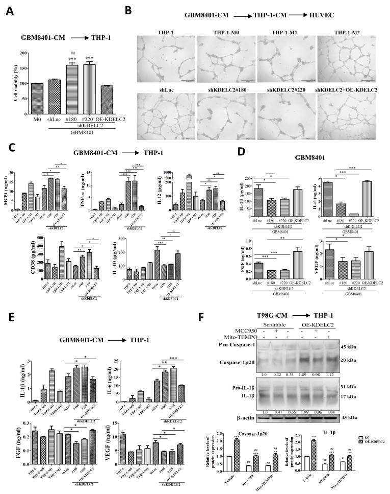
Figure 5.
KDELC2 induced the polarization of THP-1. (A) Knockdown of KDELC2 of glioblastoma cells had favorable viability of THP-1 macrophages. (B) The degree of the HUVEC proliferation of shKDELC2 and compensated OE-KDELC2 glioblastoma cells was similar to THP-1-M1 and THP-1-M2 cells, respectively. (C) High expression of MCP-1, TNF-α, IL-12, and CD38 was noted in shKDELC2 glioblastoma cells, but relatively high IL-10 expression was observed in shLuc and compensated OE-KDELC2 glioblastomas. (D) Low expression of IL-1β, IL-6, FGF, and VEGF was noted in shKDELC2 transfected glioblastoma cells. (E) High IL-1β and IL-6, but low FGF and VEGF expression in THP-1 macrophages co-cultured with shKDELC2 transfected glioblastoma-CM. (F) The application of MCC950 and Mito-TEMPO significantly increased caspase-1p20 and IL-1β in THP-1 macrophages with shKDELC2-transfected glioblastoma-CM. Bars, means ± SEM. *, p < 0.05; **, p < 0.01; ***, p < 0.001; #, p < 0.05; ##, p < 0.01; ns—non-significant.

