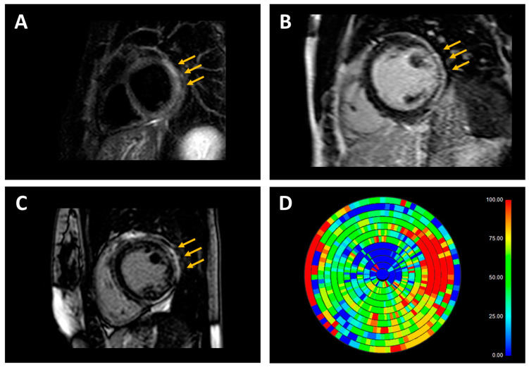
Figure 3.
Representative examples of cardiac magnetic resonance (CMR) findings in patients with inflammatory and genetic cardiomyopathies. (A) T2-weighted short-tau inversion recovery (STIR) sequence in a patient with acute myocarditis and nonischemic distribution pattern of substrate abnormalities (subepicardial hyperintensity in inferolateral left ventricular wall, arrows). (B) Late gadolinium enhancement (LGE) sequence in a patient with genetic dilated cardiomyopathy (DCM; pathogenic variant in the FLNC gene) and nonischemic distribution pattern of substrate abnormalities (subepicardial hyperintensity in inferolateral left ventricular wall, arrows). (C) LGE sequence in a patient with genetic arrhythmogenic cardiomyopathy (ACM; pathogenic variant in the DSP gene) and nonischemic distribution pattern of substrate abnormalities (subepicardial hyperintensity mainly involving the inferolateral left ventricular wall, arrows). (D) In the patient with genetic ACM, the LGE map shows an extensive left ventricular scar burden (red = maximal scar burden; blue = healthy myocardium).

