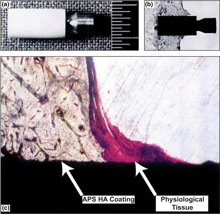
Fig. 3.
(a) Plasma-sprayed hydroxyapatite-coated implant installed in dog femur, the scale bars are 0.5 mm. (b) Histological section of an implanted zone showing the outline of the implant. (c) Zoomed-in histological section showing the integration of physiological tissue with the APS HA coating. Figure adapted from Geesink et al. (Ref 43). Used with permission of British Editorial Society of Bone & Joint Surgery, from Bonding of bone to apatite-coated implants, R.G. Geesink, K. de Groot, C.P. Klein, The Journal of Bone & Joint Surgery (British volume), volume 70, issue 1, 1988; permission conveyed though Copyright Clearance Center, Inc

