Figure 1.
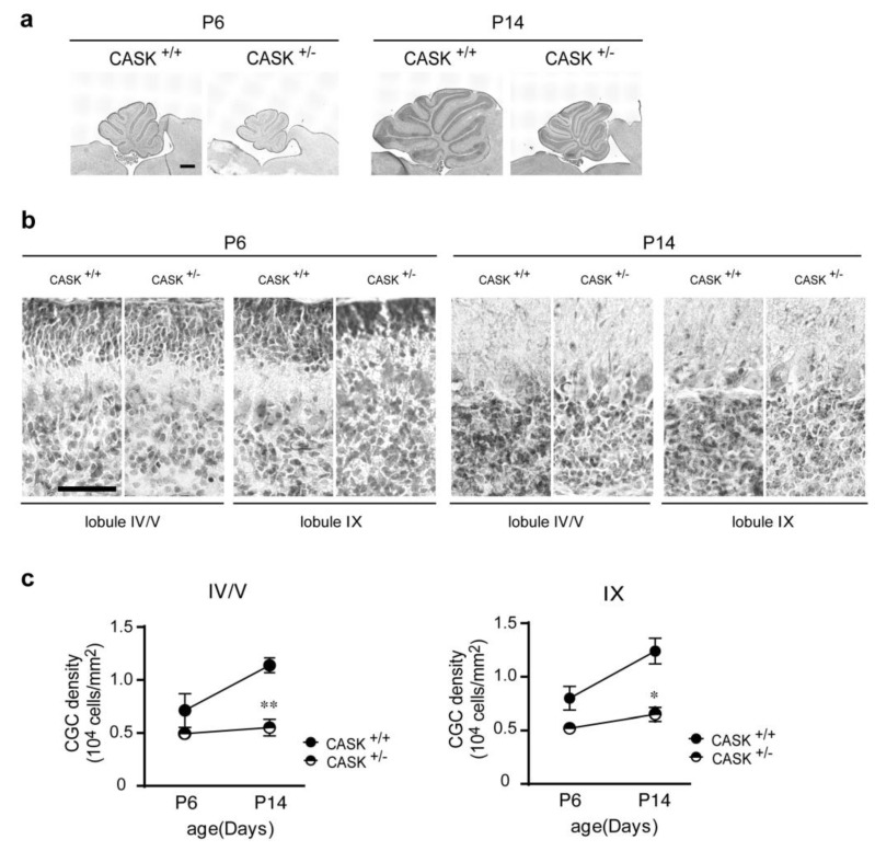
Progressive loss of CG cells in heterozygous CASK-KO mice. (a) Developmental neuroanatomical changes of the cerebella of wild-type (CASK+/+) and heterozygous CASK-KO (CASK+/−) mice. Parasagittal cerebellar sections prepared from 6- and 14-day-old wild-type and heterozygous CASK-KO mice are stained with hematoxylin. Scale bars = 500 μm. (b) Magnified images of the cerebellar cortex of 6- and 14-day-old mice in a. Lobules IV/V and IX are shown. Scale bar = 50 μm. (c) Quantification of developmental changes in CG cell densities in control and heterozygous CASK-KO mice. All values represent the mean ± s.e.m., n = 3 each from three animals. ** p < 0.01, * p < 0.05; Student t-test.

