Figure 3.
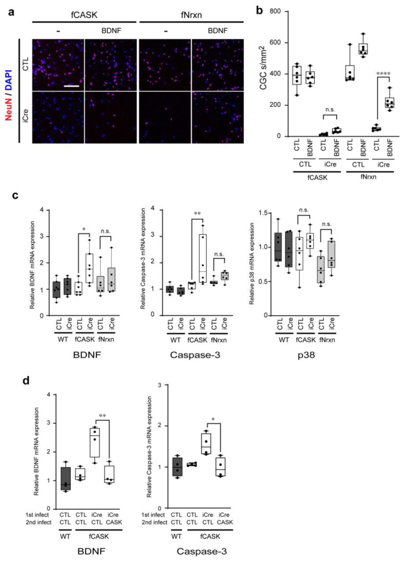
BDNF does not rescue cell survival defects in cultured CASK KO CG cells. (a) Cultured CG cells are prepared from homozygote fCASK and triple homozygote Neurexin-1, -2, -3 floxed (fNrxn) mice. BDNF is added to cultured CG cells infected with lentivirus control (CTL) or lentivirus-iCre (iCre). Cells are stained with the antibody against NeuN (red) and DAPI (blue). (b) Quantification of the number of NeuN-positive CG cells in a. Values are averages of 3 points for each well and 6 wells for each sample. **** p < 0.0001; one-way ANOVA. (c) Relative BDNF, caspase-3, and p38 expression levels in cultured CG CELLs as assessed by quantitative reverse transcriptase-PCR. Cultured CG cells are infected with lentivirus control (CTL) or lentivirus-iCre (iCre) at DIV1 and harvested at DIV5. The ratios of the mRNA expression levels of BDNF, caspase-3, or p38 to GAPDH are shown relative to that of WT CTL. All values present the mean ± s.e.m. (n = 6). (d) BDNF, and Caspase-3 mRNA expression in CASK KO CG cells rescued with lentiviral CASK infection. Cultured CG cells were infected with lentivirus control (CTL) or lentivirus-iCre (iCre) together with (CTL) or without CASK (CASK) expressing lentiviruses at DIV1 and harvested at DIV5. * p < 0.05, ** p < 0.01; one-way ANOVA, n.s. = not significant.

