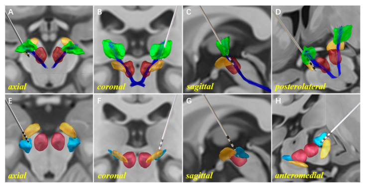
Figure 5.
An illustration of the 3D reconstruction models for unilateral Vim–PSA double-target DBS. A single electrode was implanted into the Vim–PSA double target of the left hemisphere. The 3D reconstruction of the electrode, the Vim (green), the cZi (light blue), the STN (orange), the RN (red), and the DRTT (dark blue) are shown on the axial (A,E), coronal (B,F), and sagittal (C,G) sections. For a clearer illustration of the relative positions of the electrode implanted into the Vim and PSA, perspectives are displayed from the posterolateral (D) and anteromedial (H) directions.

